Pax1/Foxa2- 1 of the primary genetic reasons why we balding men- are balding

Collapse
X
 
  • Time
  • Show
Clear All
new posts
  • eldarlmario
    Senior Member
    • Sep 2015
    • 156

    #31
    This is exciting(again- everything is IMO- and i will explain WHY it's exciting in my next post):

    It proves that we men bald men also have idiopathic scoliosis just as women who carry the pax1/foxa2 AGA/IS haplotype do- just that we have it at the http://en.wikipedia.org/wiki/Atlas_(anatomy) and for myself- i have it further down the spine as well- as in its effects extend downwards to my lower back near the http://en.wikipedia.org/wiki/Lumbar .

    I realised for a long time ago, but never thought much of- that:

    1)my right shoulder is slightly higher than my left and my right chest is bigger than my left
    2)gains from chest workouts is always more prominent on my right chest- to the point that i always do double amount of times to compensate for the left(e.g dumbell lifts for for right = X10, left = x20, pushups = right foot off the ground to focus all the body's weight on the left side to exert more resistance for my left chest)

    3)I could never stand upright 90 degrees- as in i cant do it without exerting extra effort and even so- i cant maintain it for long without experiencing constant strain. Thus, i have a natural tendency to slouch.

    Comment

    • eldarlmario
      Senior Member
      • Sep 2015
      • 156

      #32
      Malformations in the Atlas verterba could be the origin of AGA. http://en.wikipedia.org/wiki/Vertebral_artery , http://en.wikipedia.org/wiki/Dorsal_root_ganglion and http://en.wikipedia.org/wiki/Lymphatic_vessel (where Lymphocytes are carried in)

      over the entire area where the:


      over the top of the skull where it meets
      the http://en.wikipedia.org/wiki/Suture_(joint) for the http://en.wikipedia.org/wiki/Squamou...e_frontal_bone


      The upper third of the frontal bone

      Comment

      • eldarlmario
        Senior Member
        • Sep 2015
        • 156

        #33
        And i said it was exciting because(everything is IMO):

        A thorough examination of the entire http://en.wikipedia.org/wiki/Vertebral_column , especially the Atlas verterba(top of the spine) by a http://en.wikipedia.org/wiki/Neuropathology (Neurosurgeon)or http://en.wikipedia.org/wiki/Orthopedic_surgery (Orthopaedist) could even reveal some results. A correction allignment of the Atlas verterba(or https://en.wikipedia.org/wiki/Cervical_vertebrae) could even potentially 'cure' AGA by decompressing blood vessels, lymphatic vessels and nerves that transport growth factors, carries away lymphatic drainage includingwaste products and restoring ganglion cells functionality to the balding scalp and balding skull.

        Comment

        • eldarlmario
          Senior Member
          • Sep 2015
          • 156

          #34
          I have difficulty doing this exercise- turning towards my right(but not the left)- specially if i were to move my shoulders too at the same time

          Comment

          • eldarlmario
            Senior Member
            • Sep 2015
            • 156

            #35
            back to Calcitirol:

            1)decreases epidermal kerotinocyte(the skin) differentiation through inhibition of B-catenin BUT;
            2)Increases the differentiation of hair follicles

            "Deletion of VDR resulted in defects in hair differentiation in vivo, with decreased expression of ?-catenin regulated hair differentiation genes such as PADI1, hair keratin KRT31 and calcium binding protein S100a3." <===deletion of VDR = downregulation of S100a3 in hair follicles


            "Thus the major finding of this study is that while 1,25(OH)2D3(Calcitirol)/VDR inhibits the actions of B-catenin to promote keratinocyte(skin cells) proliferation, 1,25(OH)2D3/VDR promotes the ability of B-catenin to stimulate hair follicle differentiation.(inhibits B-catenin destined for the epidermal lineage BUT promotes B-catenin destined for the hair follicle lineage) This article is part of a Special Issue entitled '16th Vitamin D Workshop'."

            The active metabolite of vitamin D, 1,25-dihydroxyvitamin D3 (1,25(OH)2D3), suppresses the proliferation while promoting the differentiation of keratinocytes through the vitamin D receptor (VDR). &#946;-Catenin, on the other hand, promotes proliferation and blocks epidermal differentiation, although it s &#8230;


            Taken in context, this mean:

            Calcitriol binded VDR (UP) => thinner skin(not to be confused with fats) and increased hair growth+ S100A3

            Comment

            • eldarlmario
              Senior Member
              • Sep 2015
              • 156

              #36
              ok just found the reason why Tretinoin stops hair growth- when it will not under certain conditions- as indicated in this study:

              "We discovered that GPRC5D(this is the gene right at the top of Cotsarelis patent indicated as being regulated in haired-scalp- 19.553 folds) expression in HBC could be induced by ATRA(All-trans-Retinoid-Acid- endogenous form of Tretinoin) like other RAIG1 family genes Figure 4b (Cheng and Lotan, 1998;Brauner-Osborne and Krogsgaard-Larsen, 2000). We showed that 2 muM ATRA induced expression in a time-dependent manner (day 1, not detected; day 2, about 0.3-fold; day 3, 0.8-fold; day 4, 6.2-fold); however, the expression peaked 2 d after adding 10 muM ATRA and then decreased Figure 4b. The expression threshold was considerably higher than that in the original hair follicles (about 23-fold). The hair matrix cells (not expressing GPRC5D) are proliferating and differentiating into the cortical cells (expressing GPRC5D). When the rate of proliferation is higher than that of differentiation, the cortex continues to grow(sufficient stem cells = maintained hair grow). On the other hand, when the rate of differentiation exceeds that of proliferation, the growth was stopped because of depletion of stem cells (=matrix cells). If GPRC5D is a differentiating marker in HBC analysis, its higher expression level might indicate considerably accelerated differentiation by ATRA, the rate of which exceeds that of proliferation. A high concentration of RA is toxic( possibly why tretinoin inhibits hair growth) (Beard et al, 2001). Retinoic acids are used to treat alopecia, and are used as a depilatory(used in those minxodil-containing formulas to grow hair BUT is also used to REMOVE hair). Therefore, this transient induction may be due to excessive concentrations of ATRA and parallel the phenomena when ATRA is used as a depilatory.)"

              *So those who wants to remove their chest hair should use 10% tretinoin on their chest- on top of getting a burning itch + dry and painful skin

              Extremely low dose = gprc5d-inducing- which translates as being beneficial to hair growth. dosage used in study was 2micromolar = 600nanograms/ml in a topical. Rounded up = the nearest would be 1mcg/ml(not even 1mg), assuming we use 1ml spread all over the balding scalp, once/day.

              Generic tretinoin has 0.025% as the lowest dose. Divided by per/ml, this equates to 250mcg/ml. So difference between hair growth-inhibiting + irritating to the skin versus hair growth is 249mcg

              So basically Dosage is the critical factor that decides hair loss or hair growth.

              The problem is:

              TATA
              HNF4
              RAR <=====Retinoid acid Receptor
              RXRA
              STAT
              BATF
              COMP
              VDR
              HDAC2
              CART1
              FOXA
              FOXP1
              GATA3
              H6 family homeobox 2
              IRF
              PAX5
              P300

              For all we know, the altered RAR might be over-activated. that might translate into the fact that we have an overexpression of the RAR by ATRA(endogenous) aka Tretinoin(exogenous)- till the point that stem cells are depleted- leading to hair loss on top of causing inflammation and itch with dry skin. Which is why- Tretinoin is a very risky experimental to use- more riskier then calcitirol(which in fact by now- being inclined to overall being beneficial after going all those studies that support its role as being so) for the hair.

              Comment

              • Seuxin
                Senior Member
                • Jan 2014
                • 223

                #37
                So....what we should do ??

                Comment

                • eldarlmario
                  Senior Member
                  • Sep 2015
                  • 156

                  #38
                  Tissue and cell distribution of VDR

                  Adipose Monocytes/Macrophages
                  Adrenal Muscle, cardiac
                  Bone, osteoblasts Muscle, embryonic
                  Brain, general Muscle, smooth
                  Brain, amygdale Ovary
                  Brain, hypothalamus Pancreas ?-cell
                  Brain, glial cells Parathyroid
                  Breast Parotid
                  Cartilage Pituitary
                  Colon Placenta
                  Dendritic cells Prostate
                  Eggshell gland Retina
                  Epididymus, seminiferous tubules Skin
                  Gills (fish) Sperm
                  Hair follicle Stomach
                  Intestine Testis
                  Kidney Thymus
                  Liver Thyroid
                  Lung Tonsils, dendritic cells
                  Lymphocytes (B, T) Uterus
                  Mast cells Yolk sac

                  Comment

                  • eldarlmario
                    Senior Member
                    • Sep 2015
                    • 156

                    #39
                    CD200(upregulated by calcitriol) +CD49F aka Intergrin A6(upregulated by Tretinoin) reexpression

                    "was sufficient to reconstitute a whole follicle"- Dr Cotsarelis.

                    Comment

                    • eldarlmario
                      Senior Member
                      • Sep 2015
                      • 156

                      #40
                      ok i can see why nobody has thought of using topical calcitriol for AGA yet- there are only a few generic brands available.

                      Quote Originally Posted by princessRambo View Post
                      I am trying it, seeing small pigmented (not vellus, but very dark hair) in a 2 square inch area that was slick bald for years, and i mean slick bald, just buzzed head completely today as i am using the cream and it is difficult to apply when hair gets longer, (scalp solution seems always out of stock in that one online pharmacy), wife told me last night "you hair is getting very dark" and that made me happy [she is the actual princessRambo, her handle, when we play online games, i always thought it hilarious when she kills someone and it says: "you were killed by princessRambo" ] anyway, i think there is something to this, it has been 6 weeks so far, i will keep you posted in a couple months with pictures. I should note that I also take 3000 ml omega 3 (not fish oil amount, but actual quantity of omega 3 ingested), 6gram of evening primrose oil daily and multi vits, but i have been doing that for a while, not sure if that has interfered with the results i am seeing, i thought i would let you guys know that, as a full disclosure. Anyhow, here is a research I found noteworthy: http://www.ncbi.nlm.nih.gov/pubmed/15538745


                      This is also an interesting video, please note how he talks about injecting vd3 around 12mn into the video. I think topical cream/solution of calcipotriol can actually reach the derma papilla cell. I am thinking derma roller + calcipotriol, would that work better without inducing hypercalcemia ?
                      Really good post Princess! Keep us updated with the results!


                      this gets abit interesting

                      Comment

                      • eldarlmario
                        Senior Member
                        • Sep 2015
                        • 156

                        #41
                        Regarding FOS- information from another person(not me):

                        Definition:


                        It is involved in important cellular events, including cell proliferation, differentiation and survival; genes associated with hypoxia; and angiogenesis;[8] which makes its dysregulation an important factor for cancer development. It can also induce a loss of cell polarity and epithelial-mesenchymal transition, leading to invasive and metastatic growth in mammary epithelial cells.[9]

                        This one is higly connected with cancer.

                        The importance of c-fos in biological context has been determined by eliminating endogenous function by using anti-sense mRNA, anti-c-fos antibodies, a ribozyme that cleaves c-fos mRNA or a dominant negative mutant of c-fos. The transgenic mice thus generated are viable, demonstrating that there are c-fos dependent and independent pathways of cell proliferation, but display a range of tissue-specific developmental defects, including osteoporosis, delayed gametogenesis, lymphopenia and behavioral abnormalities.


                        Lack(and probably low level) of c-FOS leads to osteoporosis

                        It was found that overexpression of c-fos from class I MHC promoter in transgenic mice leads to the formation of osteosarcomas due to increased proliferation of osteoblasts whereas ectopic expression of the other Jun and Fos proteins does not induce any malignant tumors. Activation of the c-Fos transgene in mice results in overexpression of cyclin D1, A and E in osteoblasts and chondrocytes, both in vitro and in vivo, which might contribute to the uncontrolled growth leading to tumor. Human osteosarcomas analyzed for c-fos expression have given positive results in more than half the cases and c-fos expression has been associated with higher frequency of relapse and poor response to chemotherapy


                        Overexpression increase proliferation of osteoblasts

                        Several studies have raised the idea that c-Fos may also have tumor-suppressor activity, that it might be able to promote as well as suppress tumorigenesis. Supporting this is the observation that in ovarian carcinomas, loss of c-Fos expression correlates with disease progression. This double action could be enabled by differential protein composition of tumour cells and their environment, for example, dimerisation partners, co-activators and promoter architecture. It is possible that the tumor suppressing activity is due to a proapoptotic function. The exact mechanism by which c-Fos contributes to apoptosis is not clearly understood, but observations in human hepatocellular carcinoma cells indicate that c-Fos is a mediator of c-myc-induced cell death and might induce apoptosis through the p38 MAP kinase pathway. Fas ligand (FASLG or FasL) and the tumour necrosis factor-related apoptosis-inducing ligand (TNFSF10 or TRAIL) might reflect an additional apoptotic mechanism induced by c-Fos, as observed in a human T-cell leukaemia cell line. Another possible mechanism of c-Fos involvement in tumour suppression could be the direct regulation of BRCA1, a well established factor in familial breast and ovarian cancer.


                        This can also induce apoptosis by MAP kinase pathaway

                        L-type Ca(2+) channel activation regulates induction of c-fos transcription by hypoxia.
                        In the present study we examined the intracellular pathways that link hypoxia to activation of c-fos gene expression. Experiments were performed on rat pheocromocytoma-12 (PC-12) cells. c-fos mRNA and promoter activities were analyzed by RT-PCR and reporter gene assays, respectively. BAPTA, a Ca(2+) &#8230;


                        In the present study we examined the intracellular pathways that link hypoxia to activation of c-fos gene expression. Experiments were performed on rat pheocromocytoma-12 (PC-12) cells. c-fos mRNA and promoter activities were analyzed by RT-PCR and reporter gene assays, respectively. BAPTA, a Ca(2+) chelator, inhibited c-fos mRNA and promoter activation by hypoxia. Nitrendipine, an L-type Ca(2+)-channel blocker, abolished, whereas BAY K 8644, an L-type channel agonist, enhanced c-fos activation by hypoxia. Ca(2+) currents were augmented reversibly by hypoxia, suggesting that Ca(2+) influx mediated by L-type Ca(2+) channels is essential for c-fos activation by hypoxia. We next determined downstream pathways activated by intracellular Ca(2+) concentration. Immunoblot analysis revealed Ca(2+)/calmodulin-dependent kinase II (CaMKII) protein in PC-12 cells and revealed that hypoxia increased the enzyme activity. KN-93, a CaMK inhibitor, blocked CaMKII activation and c-fos promoter stimulation by hypoxia. Ectopic expression of an active mutant of CaMKII (pCaMKII290) stimulated c-fos promoter activity under normoxia. Hypoxia increased phosphorylation of CREB at the serine residue 133 (Ser-133), and KN-93 attenuated this effect. Point mutations at the Ca(2+)/cAMP-responsive cis-element (Ca/CRE) attenuated, whereas point mutations in the serum-responsive cis-element (SRE) abolished transcriptional activation of c-fos by hypoxia. These results demonstrate that c-fos activation by hypoxia involves CaMK activation and CREB phosphorylation at Ser-133 and requires Ca/CRE and SRE. These observations demonstrate that Ca(2+)-dependent signaling pathways play a crucial role in induction of c-fos gene expression, which may underlie long-term adaptive responses to hypoxia.


                        The promoter region of the c-fos gene is known to contain
                        specific regulatory elements that confer responsiveness to
                        phorbol esters and calcium ionophores (24-27). Thus activation
                        of protein kinase C and increases in cytosolic [Ca"] are
                        both capable of inducing c-fos expression.


                        To assess the importance of Ca2+ mobilization resulting from
                        mAChR activation, we used the Ca2+ chelator BAPTA to
                        buffer the rise in cytosolic [Ca"]. When cells are loaded with
                        20 p~ BAPTA for 30 min, carbachol no longer induces an
                        increase in cytosolic calcium (37). In BAPTA-loaded cells, the
                        mAChR-mediated increase in c-fos mRNA levels is reduced
                        by at least 75% (compare lanes 2 and 4, Fig. 5). These data
                        suggest that the increase in cytosolic [Ca'+] resulting from
                        stimulation of the mAChR is required to maximally induce cfos
                        expression.
                        In experiments using PMA and ionomycin we confirm that
                        both activation of protein kinase C and increases in intracellular
                        [Ca'+] are needed to maximally increase c-fos expression.
                        Ionomycin at a concentration of 100 nM causes a rapid and
                        transient increase in cytosolic [Ca'+] comparable with that
                        induced by carbachol (44).


                        Increase of intracellular Ca2+ -> increase in c-fos

                        Regarding BAPTA- (potential inhibitor of c-fos)

                        http://research-repository.uwa...3f7-fb6384dbc8b7).html

                        The results confirmed the relationship between EP increase and the fall of scala media CM. One interpretation of these results is that lowering the Ca2+ concentration of endolymph with BAPTA inhibits mechano-electrical transduction in outer hair cells (OHCs) and leaves the hair cell transduction channels in a closed state, thus increasing the resistance across OHCs and increasing the EP.


                        It seems that BAPTa is bad for tansduction and...

                        Effects of extracellular Ca2+ concentration on hair-bundle stiffness and gating-spring integrity in hair?cells


                        When a hair cell is stimulated by positive deflection of its hair bundle, increased tension in gating springs opens transduction channels, permitting cations to enter stereocilia and depolarize the cell. Ca2+ is thought to be required in mechanoelectrical transduction, for exposure of hair bundles to Ca2+ chelators eliminates responsiveness by disrupting tip links, filamentous interstereociliary connections that probably are the gating springs. Ca2+ also participates in adaptation to stimuli by controlling the activity of a molecular motor that sets gating-spring tension. Using a flexible glass fiber to measure hair-bundle stiffness, we investigated the effect of Ca2+ concentration on stiffness before and after the disruption of gating springs. The stiffness of intact hair bundles depended nonmonotonically on the extracellular Ca2+ concentration; the maximal stiffness of ?1200 ?N?m?1 occurred when bundles were bathed in solutions containing 250 ?M Ca2+, approximately the concentration found in frog endolymph. For cells exposed to solutions with sufficient chelator capacity to reduce the Ca2+ concentration below ?100 nM, hair-bundle stiffness fell to ?200 ?N?m?1 and no longer exhibited Ca2+-dependent changes. Because cells so treated lost mechanoelectrical transduction, we attribute the reduction in bundle stiffness to tip-link disruption. The results indicate that gating springs are not linearly elastic; instead, they stiffen with increased strain, which rises with adaptation-motor activity at the physiological extracellular Ca2+ concentration


                        and now the bad news

                        Intracellular calcium chelator BAPTA protects cells against toxic calcium overload but also alters physiological calcium responses.
                        Collatz MB1, Rüdel R, Brinkmeier H.
                        Author information
                        Abstract
                        The effect of the membrane-permeant calcium chelator 1,2-bis-(2-aminophenoxy)ethane-N,N,N',N'-tetraacetic acid tetra(acetoxymethyl) ester (BAPTA/AM) on ionomycin-induced cellular calcium overload was studied in single differentiated NH15-CA2 neuroblastoma x glioma hybrid cells. To monitor [Ca2+]i we used the fluorescent indicator Fura-2. Preincubation of the cells with 3 microM BAPTA/AM reduced the number of cells showing deregulation of [Ca2+]i during ionomycin-induced calcium influx. The calcium transients elicited by application of KCl were also severely affected by the chelator. These transients, although varying from cell to cell in shape, amplitude and duration, are well reproducible in individual cells. After incubation of cells for 1 h with 0.3-30 microM BAPTA/AM the time course of these cellular transients was markedly slowed. At 1 microM BAPTA/AM, the time constant of decline of [Ca2+]i was increased by a factor of 4.1 +/- 2.4 (n = 14) and the amplitude was reduced to about 50%. With 30 microM BAPTA/AM, the K(+)-induced calcium transients were almost completely inhibited. We conclude that intracellularly loaded calcium chelators may be used for the prevention of [Ca2+]i-induced cell damage, however, at the expense of a disturbed calcium signalling


                        In summary: I have found that BAPTA inhibit c-fos and that Ca2+ induce c-fos, unfotunetely BAPTA disturbe calcium signaling, thus BAPTA is Calcitriol antagonist - it inhibit intracelular Ca2+ :/ I ve also found that Ca2+ is crucial for mechanoelectrical tranduction of hair cells, so this is another + for calcitiol <

                        c-FOS(pro-apoptotic protein) is regulated in bald-scalp
                        high Ca2(intracellular calcium) levels => c-FOS upregulation

                        Comment

                        • eldarlmario
                          Senior Member
                          • Sep 2015
                          • 156

                          #42
                          there are tons and tons and tons and tons of generic calcitriol easily available everywhere:

                          38 pages of generic ORAL calcitirol here in the Indian generics pharmaceuticals industry


                          YET until the post anecdote linked above from ***alk, we have not head of calcitirol-induced hair regrowth in AGA-individuals anywhere- what does this tells us??

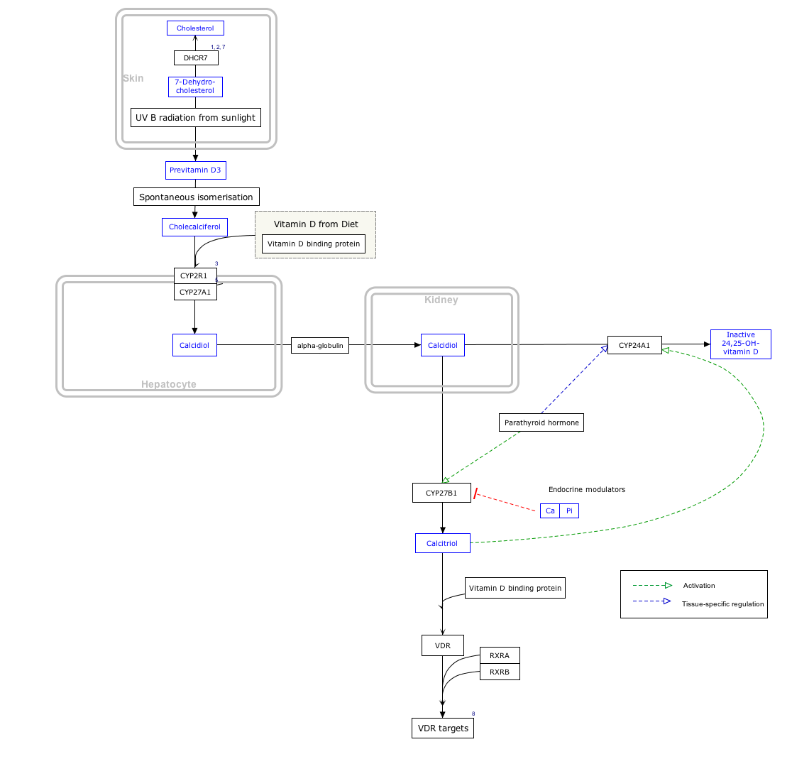
                          it proves that the hypothesis about this http://en.wikipedia.org/wiki/Vitamin_D-binding_protein might indeed be the reason why Calcitirol-binded VDR on the balding scalp is downregulated- It's not getting transported to the balding scalp/skull(but is normally expressed else where in the body) in AGA individuals.

                          Vitamin D-bind protein:

                          Comment

                          • eldarlmario
                            Senior Member
                            • Sep 2015
                            • 156

                            #43
                            "This is also an interesting video, please note how he talks about injecting vd3 around 12mn into the video. I think topical cream/solution of calcipotriol(http://en.wikipedia.org/wiki/Calcipotriol) can actually reach the derma papilla cell. I am thinking derma roller + calcipotriol, would that work better without inducing hypercalcemia ?
                            Really good post Princess! Keep us updated with the results!"

                            Calcipotriol
                            From Wikipedia, the free encyclopedia
                            Calcipotriol
                            Calcipotriol.svg
                            Systematic (IUPAC) name
                            (1R,3S,5E)-5-{2-[(1R,3aS,4Z,7aR)-1-[(2R,3E)-5-cyclopropyl-5-hydroxypent-3-en-2-yl]-7a-methyl-octahydro-1H-inden-4-ylidene]ethylidene}-4-methylidenecyclohyytyexane-1,3-diol
                            Clinical data
                            Trade names Daivobex, Dovobex, Sorilux
                            AHFS/Drugs.com monograph
                            MedlinePlus a608018
                            Pregnancy
                            category
                            AU: B3
                            US: C (Risk not ruled out)
                            Legal status
                            AU: Prescription Only (S4)
                            CA: ?-only
                            UK: Prescription-only (POM)
                            US: ?-only
                            Routes of
                            administration Topical
                            Pharmacokinetic data
                            Bioavailability 5 to 6%
                            Metabolism Hepatic
                            Excretion Biliary
                            Identifiers
                            CAS Registry Number 112965-21-6 Yes
                            ATC code D05AX02
                            PubChem CID: 5288783
                            IUPHAR/BPS 2778
                            DrugBank DB02300 Yes
                            ChemSpider 4450880 Yes
                            UNII 143NQ3779B Yes
                            KEGG D01125 Yes
                            ChEBI CHEBI:50749 Yes
                            ChEMBL CHEMBL100918
                            Chemical data
                            Formula C27H40O3
                            Molecular mass 412.605 g/mol
                            SMILES[show]
                            InChI[show]
                            (what is this?) (verify)
                            Calcipotriol (INN) or calcipotriene (USAN) is a synthetic derivative of calcitriol, a form of vitamin D. It is used in the treatment of psoriasis, marketed under the trade name "Dovonex" in the United States, "Daivonex" outside of North America, and "Psorcutan" in Germany. This medication is safe for long-term application in psoriatic skin conditions.
                            Contents [hide]
                            1 Medical uses
                            2 Adverse effects
                            2.1 Contraindications
                            2.2 Interactions
                            3 Mechanism
                            4 References
                            5 External links
                            Medical uses[edit]
                            Chronic plaque psoriasis is the chief medical use of calcipotriol.[1] It has also been used successfully in the treatment of alopecia areata.[2]

                            Adverse effects[edit]
                            Adverse effects by frequency:[1][3][4][5]
                            Very common (> 10% frequency)
                            Burning
                            Itchiness
                            Skin irritation
                            Common (1 - 10% frequency)
                            Dermatitis
                            Dry skin
                            Erythema
                            Peeling
                            Worsening of psoriasis including facial/scalp
                            Rash
                            Uncommon (0.1 - 1% frequency)
                            Exacerbation of psoriasis
                            Rare (< 0.1% frequency)
                            Allergic contact dermatitis
                            Hypercalcaemia
                            Photosensitivity
                            Changes in pigmentation
                            Skin atrophy

                            Mechanism[edit]
                            The efficacy of calcipotriol in the treatment of psoriasis was first noticed by the observation of patients receiving various forms of vitamin D in an osteoporosis study. Unexpectedly, some patients who also suffered from psoriasis experienced dramatic reductions in lesion counts.[6]
                            The precise mechanism of calcipotriol in remitting psoriasis is not well understood. However, it has been shown to have comparable affinity with calcitriol for the vitamin D receptor (VDR), while being less than 1% as active as the calcitriol in regulating calcium metabolism. The vitamin D receptor belongs to the steroid/thyroid receptor superfamily, and is found on the cells of many different tissues including the thyroid, bone, kidney, and T cells of the immune system. T cells are known to play a role in psoriasis, and it is thought that the binding of calcipotriol to the VDR modulates the T cells gene transcription of cell differentiation and proliferation related genes.


                            Normally, from my own experience of reading up information on experimental meds that might help hairloss- you would expect it's warnings/cautions section to include 'hair loss' as a potential side/adverse effect.

                            Surprisingly- Calcipotriol doesnt.

                            Comment

                            • eldarlmario
                              Senior Member
                              • Sep 2015
                              • 156

                              #44
                              ok perhaps this is why ketoconazole makes hair thin and brittle after the initial illusive-thickness-giving effect:

                              The regulation of the response of Candida albicans to hypoxic (low-oxygen) conditions is poorly understood. We used microarray and other transcriptional analyses to investigate the role of the Upc2 and Bcr1 transcription factors in controlling expression of genes involved in cell wall metabolism, er &#8230;


                              "regulation of the hypoxic response in Candida albicans.
                              Synnott JM1, Guida A, Mulhern-Haughey S, Higgins DG, Butler G.
                              Author information
                              Abstract
                              The regulation of the response of Candida albicans to hypoxic (low-oxygen) conditions is poorly understood. We used microarray and other transcriptional analyses to investigate the role of the Upc2 and Bcr1 transcription factors in controlling expression of genes involved in cell wall metabolism, ergosterol synthesis, and glycolysis during adaptation to hypoxia. Hypoxic induction of the ergosterol pathway is mimicked by treatment with sterol-lowering drugs (ketoconazole) and requires UPC2. Expression of three members of the family CFEM (common in several fungal extracellular membranes) of cell wall genes (RBT5, PGA7, and PGA10) is also induced by hypoxia and ketoconazole and requires both UPC2 and BCR1. Expression of glycolytic genes is induced by hypoxia but not by treatment with sterol-lowering drugs, whereas expression of respiratory pathway genes is repressed. However, Upc2 does not play a major role in regulating expression of genes required for central carbon metabolism. Our results indicate that regulation of gene expression in response to hypoxia in C. albicans is complex and is signaled both via lowered sterol levels and other unstudied mechanisms. We also show that induction of filamentation under hypoxic conditions requires the Ras1- and Cdc35-dependent pathway."

                              It increases c-fos expression because of high Ca2 levels in balding scalp despite inducing hypoxia- and that leads down the pro-apoptosis pathway instead of the pro-survival pathway

                              Comment

                              • eldarlmario
                                Senior Member
                                • Sep 2015
                                • 156

                                #45
                                From another person(not me)

                                Prolonged Expression of c-fos Suppresses Cell Cycle Entry of Dormant Hematopoietic Stem Cells


                                The proto-oncogene c-fos was transiently upregulated in primitive hematopoietic stem (Lin?Sca-1+) cells stimulated with stem cell factor, interleukin-3 (IL-3), and IL-6. To investigate a role of the c-fos in hematopoietic stem cells, we used bone marrow (BM) cells from transgenic mice carrying the c-fos gene under the control of the interferon-?/? - inducible Mx-promoter (Mx - c-fos), and fetal liver cells from c-fos - deficient mice. Prolonged expression of the c-fos in Lin?Sca-1+ BM cells inhibited factor-dependent colony formation and hematopoiesis on a stromal cell layer by keeping them at G0/G1 phase of the cell cycle. These Lin?Sca-1+ BM cells on a stromal layer entered into the cell cycle whenever exogenous c-fos was downregulated. However, ectopic c-fos did not perturb colony formation by Lin?Sca-1+ BM cells after they entered the cell cycle. Furthermore, endogenous c-fos is not essential to cell cycle progression of hematopoietic stem cells because the factor-dependent and the stroma-dependent hematopoiesis by Lin?Sca-1+ fetal liver cells from c-fos - deficient mice was not impaired. These results suggest that the c-fos induced in primitive hematopoietic stem cells negatively controls cell cycle progression and maintains them in a dormant state

                                The c-fos proto-oncogene, one of the immediate early genes, is transiently expressed on stimulation by external stimuli leading to cell cycle progression.10 Its product (c-Fos) forms a complex with the product of another proto-oncogene c-jun (AP-1) that regulates expression of AP-1 - binding genes at their transcriptional level.10-12 Thus, c-Fos may play a key role in the transduction of signals induced by external stimuli.12-14 c-Fos is known to be critical for the G0/G1 transition and cell cycle progression in fibroblasts.13 14The overexpression of c-fos in transgenic mice leads to a deregulated bone growth and results in sarcomas,15 16 and the overexpression in several cell lines leads to acceleration of cell cycle progression.17 18 On the contrary, overexpression of c-Fos negatively regulates cell cycle progression in some cell types.19 Thus, functions of c-Fos in cell cycle progression have remained open to question..


                                So this one definitely needs to be downregulated. However I was hoping that there wont be a need for that - c-fos is closely connected to cancer and after reading many studies - it should be balanced becouse under and overexpression leads to tumor...

                                Because prolonged expression of c-fos inhibits G0/G1 transition of dormant hematopoietic stem cells in both cytokine-dependent (Figs 2 to 5) and stroma-dependent (Fig 8) hematopoiesis, downregulation of the c-fos may initiate G0/G1 transition. Indeed, cell proliferation began in the stem cell culture from Mx - c-fos mice whenever addition of IFN-?/? to the culture was stopped (Fig 8). Therefore, c-Fos may be a gate keeper for cell cycle entry of dormant hematopoietic stem cells.


                                In summary, the c-fos was transiently induced in primitive hematopoietic stem cells stimulated with SCF, IL-3, and IL-6. The prolonged expression of c-fos inhibited cell-cycle entry of primitive hematopoietic stem cells stimulated with SCF, IL-3, and IL-6 as well as in case of cultures on a stromal cell layer. Hematopoietic stem cells with the c-fos expression in culture survived in a dormant state and entered the cell cycle after c-fos was downregulated. We propose that c-Fos plays the role of gate keeper in cell cycle progression of dormant hematopoietic stem cells.

                                Comment

                                Working...